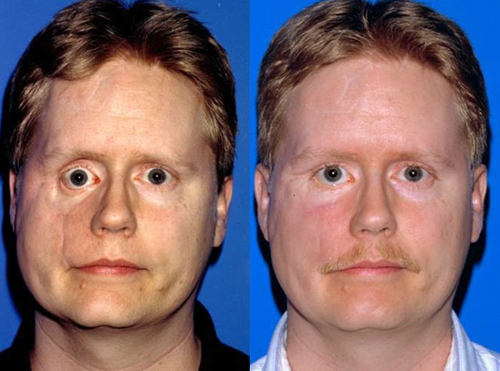
Enophthalmos: Understanding the Sunken Eye Condition
Introduction
Enophthalmos is a medical condition characterized by the posterior displacement of the eyeball within the orbit, resulting in a sunken appearance of the eye. This condition can have various etiologies and may present with a range of symptoms that necessitate thorough clinical evaluation. Understanding enophthalmos is crucial for both healthcare professionals and patients, as it can lead to functional and aesthetic concerns. This article will explore the historical background, anatomy and pathophysiology, causes, symptoms and clinical presentation, diagnosis, treatment options, prognosis and recovery, living with enophthalmos, current research, and future directions.
What is Enophthalmos?
Enophthalmos refers to the sinking of the eyeball into the orbital cavity. It is the opposite of exophthalmos, where the eye protrudes outward. Enophthalmos can be congenital or acquired and may result from trauma, diseases, or surgical procedures. The condition can affect one or both eyes and may lead to significant functional impairments and cosmetic concerns for the patient.
Historical Background
The understanding of enophthalmos has evolved over time. Historically, this condition was often overlooked or misdiagnosed due to a lack of advanced imaging techniques. Early descriptions of enophthalmos date back to ancient medical texts that noted variations in eye positioning but did not fully understand the underlying causes.In modern medicine, advancements in imaging technologies such as computed tomography (CT) scans and magnetic resonance imaging (MRI) have allowed for better diagnosis and understanding of enophthalmos. Research has focused on identifying various causes, including trauma and disease processes that lead to this condition.
Anatomy and Pathophysiology
To understand enophthalmos fully, it is essential to grasp the anatomy involved:
- Orbit: The bony structure surrounding the eye that houses the eyeball and associated tissues.
- Extraocular Muscles: These muscles control eye movement and help maintain proper positioning within the orbit.
- Fat Pads: Orbital fat helps cushion the eye and maintain its position.
In cases of enophthalmos:
- Orbital Volume Loss: The volume of the orbit may decrease due to factors such as trauma or disease processes.
- Displacement: The eyeball may be displaced posteriorly into the orbit due to structural changes or loss of supporting tissues.
Understanding these anatomical components is crucial for diagnosing and treating enophthalmos effectively.
Causes
Several factors contribute to the development of enophthalmos:
- Traumatic Causes: Orbital fractures, particularly blow-out fractures, can result in displacement of orbital contents, leading to enophthalmos. Trauma can cause immediate or gradual displacement as healing occurs.
- Inflammatory Causes: Conditions such as orbital cellulitis or chronic sinusitis can lead to inflammation that affects orbital structure and position.
- Neoplastic Causes: Tumors within or around the orbit can exert pressure on the eyeball, causing it to sink back into the orbit.
- Surgical Causes: Certain surgical procedures involving the orbit may inadvertently lead to changes in eye positioning.
Understanding these causes is crucial for implementing effective prevention strategies and appropriate treatment options.
Symptoms and Clinical Presentation
The symptoms of enophthalmos may vary depending on its underlying cause but commonly include:
- Sunken Appearance: A noticeable difference in eye position compared to the other eye.
- Diplopia: Double vision may occur due to misalignment caused by extraocular muscle involvement.
- Visual Changes: Some patients may experience blurred vision or other visual disturbances.
- Eyelid Changes: Associated conditions may lead to eyelid retraction or ptosis (drooping).
Recognizing these symptoms early can facilitate timely medical intervention.
Diagnosis
Diagnosing enophthalmos involves several steps:
- Medical History Review: A thorough history including any recent trauma, surgeries, or underlying health conditions.
- Physical Examination: An ophthalmologist will assess ocular motility, visual acuity, and symmetry between both eyes.
- Imaging Studies:
- CT Scan: Provides detailed images of bony structures around the eye; useful for identifying fractures or tumors.
- MRI: Useful for assessing soft tissue structures within the orbit.
- Objective Measurements: Techniques such as using a ruler or specialized instruments can quantify the degree of enophthalmos by measuring differences in globe position.
Early diagnosis is critical for effective management and reducing risks associated with untreated enophthalmos.
Treatment Options
Treatment for enophthalmos depends on its underlying cause:
- Medical Management:
- Treating underlying infections with antibiotics if inflammation or infection is present.
- Managing chronic conditions that contribute to orbital changes.
- Surgical Interventions:
- Surgical correction may involve fat transfer or soft tissue surgery to restore normal eye position.
- Orbital reconstruction may be necessary in cases involving significant trauma or structural abnormalities.
Regular monitoring through follow-up appointments is essential to assess treatment efficacy and detect any recurrence early.
Prognosis and Recovery
The prognosis for individuals with enophthalmos varies based on several factors:
- Timeliness of Treatment: Early intervention significantly improves outcomes; most patients respond well when treated promptly.
- Severity of Condition: Patients with mild forms may respond well to medical management while those with severe forms might require more aggressive surgical interventions.
After successful treatment, many individuals can expect an improvement in their symptoms; however, ongoing monitoring remains crucial due to potential recurrence or complications.
Living with Enophthalmos
Living with enophthalmos requires proactive health management:
- Regular Check-ups: Individuals should maintain regular appointments with their healthcare provider for monitoring overall health.
- Lifestyle Modifications:
- Protecting eyes from injury
- Managing stress
- Engaging in regular physical activity
These lifestyle changes can help manage symptoms and reduce recurrence risks. Emotional support from friends or support groups can also be beneficial as individuals navigate their diagnosis and treatment options.
Research and Future Directions
Current research efforts focus on improving understanding and management strategies for enophthalmos:
- Innovative Treatments: Ongoing studies explore new surgical techniques that could provide more effective management with fewer side effects.
- Genetic Studies: Investigating genetic predispositions could lead to better prevention strategies for at-risk populations.
Continued research will enhance clinical practices surrounding this condition while improving patient outcomes in future years.
Conclusion
Enophthalmos is a significant condition that requires careful attention due to its potential implications for ocular health. With advancements in diagnostic techniques and treatment modalities available today, many individuals can manage this condition effectively. Increased awareness among healthcare providers about risk factors, types of enophthalmos, and appropriate management strategies is essential for improving patient care in this area.
Disclaimer: This article is intended for informational purposes only and should not be considered medical advice. Always consult a healthcare professional for diagnosis and treatment options.

