Tinea Capitis: Understanding, Diagnosis, and Treatment of Scalp Ringworm

Introduction
Tinea capitis, commonly known as scalp ringworm, is a fungal infection that affects the scalp and hair shafts. This condition is particularly prevalent among children but can affect individuals of any age. Understanding tinea capitis is essential for early diagnosis and effective treatment, as it can lead to hair loss and significant discomfort if left untreated. This article will provide a comprehensive overview of tinea capitis, including its causes, symptoms, diagnosis, treatment options, and preventive measures.
What Is Tinea Capitis?
Tinea capitis is a dermatophyte infection caused by fungi that invade the outer layer of the skin on the scalp. The most common pathogens responsible for this infection include:
- Trichophyton tonsurans: The leading cause of tinea capitis in the United States.
- Microsporum canis: Often transmitted from pets to humans.
- Trichophyton violaceum: More common in certain geographical areas.
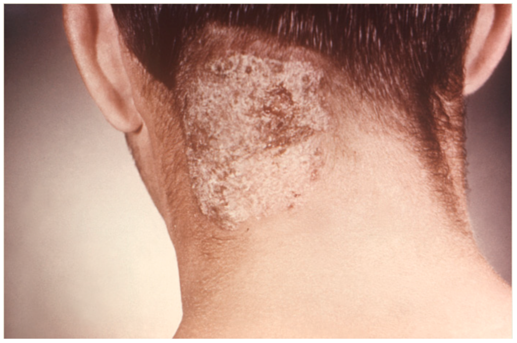
The infection typically presents as scaly patches on the scalp that may become inflamed and result in hair loss. Tinea capitis is contagious and can spread through direct contact with an infected person or indirectly through shared items such as combs, hats, or towels.
Historical Background
The history of tinea capitis dates back centuries, with early descriptions of fungal infections affecting the scalp appearing in medical texts. In the late 19th century, dermatologists began to classify various skin infections caused by fungi, leading to a better understanding of tinea capitis. The term “ringworm” was used due to the characteristic ring-like appearance of the lesions.As research progressed, scientists identified specific fungal species responsible for tinea capitis and developed more effective treatments. Today, awareness of this condition has increased among healthcare providers and the public, leading to improved diagnostic techniques and management strategies.
Anatomy and Pathophysiology
The anatomy involved in tinea capitis includes:
- Scalp Skin: The outer layer of skin on the head where the infection occurs.
- Hair Follicles: Structures from which hair grows; these can become infected by dermatophytes.
The pathophysiology of tinea capitis involves the invasion of dermatophyte fungi into the stratum corneum (the outermost layer of skin) and hair follicles. Once the fungi invade these structures, they begin to multiply, leading to inflammation and damage. The immune response may also contribute to symptoms such as redness, swelling, and itching.
Causes
Tinea capitis is primarily caused by dermatophyte fungi. Factors contributing to its development include:
- Direct Contact: Coming into contact with an infected person or animal can lead to transmission.
- Shared Items: Using contaminated personal items like combs, hats, or towels increases risk.
- Environmental Factors: Fungi thrive in warm, humid environments; overcrowded living conditions may facilitate spread.
- Weakened Immune System: Individuals with compromised immune systems are more susceptible to fungal infections.
Symptoms and Clinical Presentation
Symptoms of tinea capitis can vary based on the individual and the specific fungal species involved. Common signs include:
- Scaly Patches: Round or oval patches on the scalp that may be dry or scaly.
- Hair Loss: Patches of hair loss may occur as hair shafts break off at the scalp surface.
- Itching: The affected area often becomes itchy and uncomfortable.
- Inflammation: Redness and swelling may develop around infected patches.
- Kerion Formation: In some cases, a kerion (an inflamed mass) may form, resulting in pus-filled lesions that can lead to scarring.
In severe cases or with specific fungal types (e.g., Microsporum canis), additional symptoms such as fever or swollen lymph nodes may occur.
Diagnosis
Diagnosing tinea capitis typically involves several steps:
- Medical History: A thorough review of symptoms, exposure history (e.g., contact with infected individuals), and any previous skin conditions is essential.
- Physical Examination: Healthcare providers will inspect the scalp for characteristic lesions and signs of inflammation.
- Laboratory Tests:
- KOH Preparation: A sample of hair or skin scrapings is treated with potassium hydroxide (KOH) to visualize fungal elements under a microscope.
- Fungal Culture: Culturing samples can help identify specific fungal species responsible for the infection.
- Wood’s Lamp Examination: Some fungi fluoresce under ultraviolet light; this test can help identify certain types of infections.
Treatment Options
Treatment for tinea capitis focuses on eradicating the fungal infection and alleviating symptoms:
Medical Treatments
- Oral Antifungal Medications:
- Griseofulvin is commonly prescribed for 4 to 8 weeks as it effectively targets dermatophyte infections.
- Terbinafine (Lamisil) is another effective option that may be preferred due to its shorter treatment duration.
- Topical Antifungals:
- While topical treatments alone are generally insufficient for treating tinea capitis, they may be used in conjunction with oral medications to prevent spread or reinfection.
- Corticosteroids:
- For severe inflammation or kerion formation, short courses of corticosteroids may be prescribed to reduce swelling and discomfort.
Home Remedies
- Medicated Shampoos:
- Antifungal shampoos containing ketoconazole or selenium sulfide can help reduce fungal load on the scalp but should not replace systemic antifungal treatment.
- Hygiene Practices:
- Regularly washing bed linens, hats, and brushes with hot water can help prevent reinfection.
- Avoid sharing personal items such as combs or hats.
Prognosis and Recovery
The prognosis for individuals with tinea capitis is generally favorable with appropriate treatment:
- Most patients respond well to oral antifungal medications; symptoms typically improve within weeks.
- It is crucial to complete the full course of treatment to prevent recurrence or chronic infection.
- Follow-up appointments may be necessary to ensure complete resolution of infection.
Living with Tinea Capitis
Managing life with tinea capitis involves several considerations:
- Regular Monitoring: Consistent follow-up appointments with healthcare providers are crucial for managing ongoing symptoms effectively.
- Educating Yourself: Understanding your condition helps you make informed decisions about your health care.
- Support Systems: Engaging with support groups can provide emotional support from others facing similar challenges.
- Healthy Lifestyle Choices: Maintaining a balanced diet rich in nutrients can support overall well-being during recovery.
Research and Future Directions
Ongoing research into tinea capitis focuses on understanding its underlying mechanisms better and developing innovative strategies for prevention:
- Vaccine Development: Researchers are exploring potential vaccines against common dermatophytes responsible for tinea capitis.
- Genetic Studies: Investigating genetic factors that influence susceptibility could lead to personalized prevention strategies.
- Public Awareness Campaigns: Increased awareness through educational campaigns aims to reduce incidence rates by informing communities about prevention techniques.
Conclusion
Tinea capitis is a common yet manageable condition that requires awareness for effective treatment. Understanding its causes, symptoms, diagnosis methods, treatment options, and lifestyle adjustments can empower individuals facing this condition. If you suspect you have tinea capitis or experience persistent symptoms related to your scalp health, seeking medical advice is crucial for appropriate care.
Disclaimer
This article is intended for informational purposes only and should not be considered medical advice. Always consult a healthcare professional for diagnosis and treatment options tailored to your specific needs.
